Artículo original
Vol. 46 Núm. 1 (2026)
36 min
Main characteristics of oesophagitis as food allergy component: From aetiology to prevention
María Zofia LISIECKA
Instituto Médico Nacional del Ministerio del Interior y de la Administración.
*Correspondencia:
María Zofia Lisiecka mariazofialisiecka@gmail.com
Recepción:
22/09/2025
Aceptación:
22/02/2026
Publicación:
03/03/2026
Cómo citar
María Zofia LISIECKA. Main characteristics of oesophagitis as food allergy component: From aetiology to prevention. Nutr Clín Diet Hosp. 2026; 46(1):307-317 DOI: 10.12873/461lisiecka
Métricas
599
Visualizaciones
85
Descargas
Compartir
Compartir
Resumen (EN)
Introduction: Eosinophilic oesophagitis is a chronic, Th2-mediated condition characterised by symptoms of oesophageal dysfunction and histological evidence of eosinophils in inflammatory infiltrates.
Aim: The narrative review with structured search aims to analyse the relationship between eosinophilic oesophagitis and food allergy.
Materials and Methods: The structured search for pertinent publications containing essential information on eosinophilic oesophagitis published in peer-reviewed journals in Polish, French, German, and English from 2017 to 2024 was conducted in PubMed, ResearchGate, Scopus, Web of Science and Google Scholar databases.
Results: Symptoms of eosinophilic oesophagitis vary between children and adults. The incidence of cases is 5-30 per 100,000 people, depending on the region, and the prevalence is 0.5-1 case per year. The pathogenesis is currently being studied, but the allergic nature of eosinophilic oesophagitis is noted: the presence of concomitant allergic diseases; peripheral eosinophilia; elevated levels of total immunoglobulin E (IgE); the presence of specific immunoglobulins to food and inhalation allergens in patients; reduction of symptoms after elimination diet and steroids. The most used diagnostic methods are biopsy of oesophageal samples, endoscopic ultrasound, oesophageal thread test, and gene expression studies. New tools are being developed for early diagnosis, as timely diagnosis significantly reduces the risk of severe complications. For treatment purposes, dietary nutrition (elimination, elementary and empirical diets), proton pump inhibitors, and local and systemic steroids are used, and in case of significant narrowing of the oesophagus, its dilation is performed. Highly effective medicines based on monoclonal antibodies are being developed to improve the quality of life of patients and stop the progression of the disease.
Conclusion: Thus, eosinophilic oesophagitis is a relatively uncommon disease with an allergic nature that requires further study to develop effective diagnostic and treatment methods.
Palabras clave (EN):
Th2 immunity
hypersensitivity
oesophageal biopsy
eosinophilic inflammation
dysphagia
INTRODUCTION
The incidence of eosinophilic oesophagitis may be partially attributed to the decline in food quality, water contamination, and air pollution. The age of manifestation of the disease varies from several months to years. Eosinophilic oesophagitis can be of immunological origin, caused by ingested or inhaled allergens. Risk factors include a family history of allergic diseases, antibiotic use at an early age, and the presence of atopic diseases. Caucasian men are three times more likely to be affected than women. Pathology can occur at any age and manifest itself with a spectrum of different symptoms. Children are more likely to develop acute inflammation, and in adults, the clinical picture is caused by fibrosis. In infants, nonspecific symptoms of oesophageal lesions, such as vomiting, refusal to eat, and growth and developmental delays, have been described. In young and middle-aged people, pain when swallowing, difficulty swallowing and narrowing of the oesophagus are observed. It is characteristic that the symptoms of the disease differ between men and women. However, it should be noted that chest pain and heartburn predominate in women, and swallowing difficulties in men.
The problem is late diagnosis. This is due to non-specific symptoms and the lack of specific markers of the disease1. The exact causes of the disease are unknown and are under investigation. Polish scientists Kanikowska et al.2, Savarino and Barbara3, Wojas et al.4 described two peaks in the incidence: the first at the age of 5-11 and the second at the age of 35-55. The exact causes and mechanism of atopic dermatitis are unknown, but these scientists believe that the disease is associated with immune, genetic and environmental factors. Muir and Falk5 noted that each year of untreated eosinophilic oesophagitis increased the risk of strictures by 9%. In patients with a disease duration of more than 21 years, the incidence of strictures was 50%, and dysphagia – 60%. Zhang et al.6 proposed the oesophageal microbiome as a biomarker of eosinophilic oesophagitis. The study revealed that patients with eosinophilic oesophagitis have significantly more diverse bacterial populations in their oesophagus compared to those with gastroesophageal reflux disease. While analysing oesophageal bacterial content shows promise as a tool for early diagnosis and differentiation of eosinophilic oesophagitis, this diagnostic approach remains investigational. Pytrus et al.7 addressed the possibility of widespread use of endoscopic ultrasound in eosinophilic oesophagitis. This method of diagnosis is promising but is currently rarely used in clinical practice. Polish scientists Sarbinowska et al.8 addressed predictors of non-invasive diagnosis of eosinophilic oesophagitis, namely, the relationship between the level of interleukin 5, transforming growth factor, basic protein and eotaxin 3 in plasma and the severity of eosinophilic oesophagitis. At the same time, the disadvantage of this study is the small sample of participants, which cannot guarantee similar results in the population. The search for an etiological treatment is still under development. Kuźmiński et al.9 concurrently suggested efficacious therapeutic alternatives that alleviate symptoms and enhance the quality of life for patients. Topical steroids are the first-line therapy, but prolonged interaction with the oesophageal mucosa is required to increase their efficacy and histological improvement. Główczewski and Krogulska10 described new medicinal products for the delivery of topical corticosteroids.
Eosinophilic oesophagitis remains a complex condition with ongoing debates surrounding its causes, early detection, and optimal treatment approaches. This study focuses on examining the relationship between eosinophilic oesophagitis and food allergies to better understand this inflammatory disorder. The research seeks to clarify the underlying causes and risk factors that contribute to disease development, while exploring the mechanisms by which the condition affects oesophageal tissue. Additionally, the study will examine current diagnostic approaches and treatment strategies, with particular attention to how allergic hypersensitivity influences disease progression. Through comprehensive analysis of clinical presentations and therapeutic outcomes, this work aims to identify practical lifestyle modifications that patients can implement to enhance their daily functioning and overall well-being while managing eosinophilic oesophagitis.
MATERIALS AND METHODS
The study is a narrative review with structured search for relevant information on eosinophilic oesophagitis which was conducted in PubMed, ResearchGate, Scopus, Web of Science and Google Scholar databases in the first stage of the study, in September 2024.
All pertinent publications containing essential information (clinical trials, randomised controlled trials, reviews, systematic reviews, and meta-analyses) published in peer-reviewed journals in Polish, French, German, and English from 2017 to 2024 were identified and evaluated. Search for the required information using a combined set of keywords: “allergic reaction”, “immune response”, “hypersensitivity”, “cross-reaction”, “food allergy”, “oesophageal inflammation”, “dysphagia”, “oesophageal stenosis”, “eosinophilic oesophagitis”, “non-IgE-mediated food allergy”, “epidemiology of eosinophilic oesophagitis”, “aetiology of eosinophilic oesophagitis”, “symptoms of eosinophilic oesophagitis”, “mechanism and diagnosis of development of eosinophilic oesophagitis”, “treatment and prevention of eosinophilic oesophagitis”, “complications of eosinophilic oesophagitis”, “elementary diet”, “empirical diet”, “elimination diet”.
To ensure the inclusion of relevant and high-quality articles, a comprehensive search equation was applied. The search equation for PubMed included Boolean operators (AND, OR) and MeSH terms to combine multiple key concepts: (“allergic reaction” OR “immune response” OR “hypersensitivity” OR “cross-reaction”) AND (“food allergy” OR “oesophageal inflammation” OR “eosinophilic oesophagitis” OR “dysphagia” OR “oesophageal stenosis”) AND (“epidemiology” OR “aetiology” OR “symptoms” OR “diagnosis” OR “treatment”) AND (“2017”[Date - Publication]: “2024”[Date - Publication]).
The initial search yielded 1460 entries, which underwent systematic evaluation through title and abstract screening to assess relevance, publication timeline, and evidence quality. Reference lists of relevant articles were examined to identify additional sources. Duplicate studies and those outside the specified publication timeframe were excluded, followed by application of predetermined inclusion and exclusion criteria. The inclusion criteria comprised clinical studies such as clinical trials, randomized controlled trials, reviews, systematic reviews, and meta-analyses, involving human populations with eosinophilic oesophagitis. The inclusion of heterogeneous study designs aimed to provide a comprehensive overview of eosinophilic oesophagitis. A narrative synthesis approach was used, prioritising findings from clinical trials and RCTs for their clinical relevance, while using reviews and meta-analyses to identify broader trends. The synthesis was guided by the level of evidence and methodological rigor to ensure reliable conclusions. Studies were required to address outcomes related to the epidemiology, aetiology, risk factors, pathogenesis, clinical presentation, diagnostic methods, treatment approaches, or prevention strategies of eosinophilic oesophagitis. Only peer-reviewed articles published in English, Polish, French, or German between 2017 and 2024 were included. The review focused on papers providing relevant information about eosinophilic oesophagitis epidemiology, aetiology, risk factors, developmental mechanisms, clinical presentation, diagnostic methods, treatment approaches, and prevention strategies. Studies examining the pathophysiological connections of eosinophilic oesophagitis and the effects of proton pump inhibitors and corticosteroids were included. The exclusion criteria eliminated animal studies, non-peer-reviewed content, and studies published before 2017. Articles focusing on unrelated allergic diseases or non-allergic conditions were also excluded, as well as those that lacked scientific rigor (those with insufficient sample sizes, methodological flaws, or unverified data.) or provided outdated clinical information (studies published more than seven years prior to the review date).
The screening and selection process was conducted by two reviewers who assessed the relevance and quality of the studies based on the predefined criteria. The initial search yielded a large number of articles, which were filtered using title and abstract screening, followed by full-text evaluation. Discrepancies, if any, would have been resolved by consensus. This approach minimised potential biases by applying consistent and explicit inclusion and exclusion criteria, ensuring that only relevant, high-quality studies were selected for inclusion in the review.
The selection encompassed research on various allergy types and novel approaches to studying allergic reactions, without restrictions based on gender, age, race, geographic location, social status, or disease characteristics. Studies describing non-IgE-mediated allergic reactions were incorporated, along with research on different dietary interventions including elementary, elimination, and empirical diets. This systematic selection process ultimately identified 50 articles that fully satisfied the established criteria. To ensure accuracy, selected sources underwent re-evaluation, processing, and verification. All cited studies comply with technical requirements and copyright standards.
RESULTS AND DISCUSSION
Risk factors and aetiology of eosinophilic oesophagitis
Eosinophilic oesophagitis is a chronic Th2-mediated condition characterised by clinical manifestations of oesophageal dysfunction and a histological prevalence of eosinophils in inflammatory infiltrates. The incidence is 5-30 cases per 100,000 people, depending on the region, and the prevalence is 0.5-1 case per year. The prevalence is higher in North America and Western countries compared to Asian countries. Men are 1.5-3 times more likely to be affected than women. The percentage of adults to children is approximately 1.2:111. The contributing environmental factors include prematurity, caesarean section, newborns staying in intensive care units, antibiotics at an early age, feeding with artificial formula, creating “sterile” conditions and lack of contact with microbes, and high-fat content in the diet. Infectious diseases can contribute to the development of eosinophilic oesophagitis. Kanikowska et al.2 described that Herpes Simplex can damage the oesophageal mucosa and promote its eosinophilic inflammation. Eosinophilic oesophagitis may result from diminished expression of the CCL26 gene, responsible for encoding the chemokine eotaxin-3, which facilitates the migration of eosinophils to the oesophagus. Irregularities in the expression of desmoglein-1, calpain-14, and filaggrin also play a role in the disease’s progression. A history of allergic diseases increases the risk of developing eosinophilic oesophagitis. If one of the siblings has eosinophilic oesophagitis, the risk of contracting the disease increases by 2.5 times. Eosinophilic oesophagitis often develops against the background of genetic diseases. In connective tissue diseases (Loeys-Dietz and Ehlers-Danlos syndrome), there is an increase in TGF-b production, which leads to an increase in the contractile function of smooth muscles and tissue remodelling. Eosinophilic oesophagitis is linked to Netherton syndrome, which results from autosomal dominant mutations in the protease inhibitor SPINK5. Eosinophilic oesophagitis is linked to autoimmune disorders, including Hashimoto’s thyroiditis, rheumatoid arthritis, coeliac disease, and multiple sclerosis.
Under physiological conditions, the cells of the oesophagus do not contain eosinophils. The action of damaging factors results in the release of cytokines interleukin 5, 25 and 33. This triggers the activation of immune cells: natural killer T cells, CD4+ T helper 2 memory cells, and type 2 innate lymphoid cells. Interleukin 5 causes the proliferation and spread of eosinophils into the peripheral blood and further into the oesophageal wall12-14. Most often, Chang et al.12 note the main role of food and inhalation antigens in the pathogenesis of eosinophilic oesophagitis. This is confirmed by the fact that patients achieve remission when allergenic foods are eliminated, and when trigger foods are reintroduced, the disease relapses. It is also necessary to maintain the integrity of the oesophageal epithelial barrier. If its permeability is compromised, the basal zone proliferates and the intercellular gaps in the suprabasal layers enlarge. Transmural inflammation of the oesophagus wall induces fast remodelling, resulting in lamina propria fibrosis, basal cell hyperplasia, and smooth muscle hypertrophy. Study Wojas et al.4 indicate the influence of the oesophageal microbiome on the development of eosinophilic oesophagitis. Thus, in this disease, the number of Haemophilus increases and Firmicutes decreases. The number of Neisseria and Corynebacterium increased in the affected oesophageal mucosa in children, while Streptococcus and Atopobium were found in healthy children.
Pathophysiology of eosinophilic oesophagitis and pathogenesis of dysphagia
Under normal conditions, the oesophageal epithelium is presented as a non-cornified squamous multilayered epithelium. It is constructed of 3 layers: basal, suprabasal and horny. The first layer consists of proliferative cells with a total thickness of up to 15% of the epithelium. The oesophageal mucosa consists of multiple layers, with the second containing transitional basal cells and the third comprising simple and multilayered squamous epithelial cells. This multilayered squamous non-keratinising epithelium, along with its protective mucus layer, shields underlying tissues from microbial, mechanical, and chemical damage. In eosinophilic oesophagitis, epithelial barrier dysfunction occurs with reduced transepithelial resistance. Three key factors drive these changes: the epidermal differentiation complex (EDC), filaggrin, and Calpain 14. The EDC represents a gene cluster at chromosome locus 1q21 that regulates epithelial differentiation. Filaggrin produces an intracellular protein that aggregates keratin filaments while simultaneously decreasing essential junction proteins including E-cadherin, claudin-1 and 7, zonulin-1, and desmoglein-1. Calpain 14, an intracellular regulatory protease specific to oesophageal mucosa, maintains epithelial barrier integrity but becomes significantly elevated during eosinophilic oesophagitis, contributing to barrier compromise.
In eosinophilic oesophagitis, the basal zone of the oesophagus is hypertrophied, with the basal cell layer constituting more than 15% of the overall epithelial thickness. The exact mechanisms are not known, but it has been described that the calcium-activated anoctamin chloride channel regulates chloride production and movement in oesophageal epithelial cells, resulting in abnormal proliferation. This occurs by phosphorylating cyclin-dependent kinase 2 and passing through the G1/S cell cycle checkpoint. An increased amount of anoctamin chloride channel 1 m ribonucleotides was detected in oesophageal biopsies of patients, and its level correlated with the thickness of the basal zone. At endotype 3, anoctamin chloride channel 1 exposure is associated with the formation of fibrostenosis by increasing chronic inflammation and developing severe histological changes15-17.
The expansion of intercellular spaces correlates with the severity of eosinophilic oesophagitis. The exact mechanisms of this pathological process are unknown, but it is associated with the influence of the solute carrier family 9, subfamily A, 3 (SLC9A3). The influence of acid on intercellular spaces results in the establishment of an electrochemical gradient and the passage of chloride ions, which generates an osmotic force facilitating water movement and the expansion of intercellular gaps. The extent of SLC9A3/NHE3 m ribonucleotides release correlates with the number of eosinophils and the degree of intercellular space expansion. A decrease in the size of intercellular spaces is associated with a decrease in clinical manifestations of the disease18-20.
The primary histology indicator of the disease is the presence of intraepithelial eosinophils within the oesophagus. Their number correlates with the severity of the disease. As such, Cavalli et al.21 and O’Shea et al.22 described those patients with severe oesophageal eosinophilia have a more severe course (clinically, histologically and endoscopically) compared to those with low-grade oesophageal eosinophilia. This is partly explained by the degranulation of eosinophils, the release of several cytotoxic substances (eosinophilic peroxidase, basic protein, eosinophilic cationic protein and eosinophilic neurotoxin), and their deposition in the extracellular space. The presence of these substances correlates with the severity of the disease. The effect of neutralising monoclonal antibodies reduced the level of eosinophilia in the oesophagus and slowed angiogenesis and fibronectin deposition. This indicates a significant impact of eosinophils on the pathogenesis of the disease.
In patients with exacerbation of eosinophilic oesophagitis, CCL26 m ribonucleotides is increased in biopsy samples. This family of chemokines is responsible for the transport of eosinophils, and when the normal functioning of the CCR3 gene is impaired, allergen-induced eosinophilic inflammation of the oesophagus develops. Eosinophilic oesophagitis is also associated with decreased production of genes responsible for the barrier function of the oesophagus (in particular, SPRR, desmoglein 1 and involutionrin). The genes SPINK5 and SPINK7 are also reduced, which contributes to the loss of protective function of the oesophageal wall and increased expression of urokinase activator (which promotes eosinophil activation). Blood samples from patients show elevated levels of CD4 T cells, the number of which correlates with eosinophilia in the oesophagus. These cells are responsible for the expression of IL-4, IL-5 and IL-13. It is worth noting that as a result of glucocorticoid therapy, the number of IL-4 decreases, which also confirms the allergic aetiology of eosinophilic oesophagitis.
An elevation in mast cell quantity and evidence of degranulation were seen in oesophageal biopsy specimens. Bolton et al.23 reported an elevation in mast cell quantity in patients exhibiting endoscopic and clinical manifestations. These cells penetrate all layers of the oesophagus and impair muscle contractility. TGF-β1 levels are elevated in tryptase-positive mast cells; this cytokine stimulates an increase in the number of smooth muscle cells and contributes to the construction of the oesophagus. Scientists Khokhar et al.15 demonstrated an increase in the level of total and food-specific serum IgG4 and its deposition in the oesophageal tissues. Periostin is an extracellular matrix protein synthesised by fibroblasts and oesophageal epithelial cells in response to TGF-β and IL-13, facilitating eosinophil adherence and migration while regulating extracellular matrix deposition. Its expression is elevated 40-50 fold in biopsies, but not in the serum of patients with eosinophilic oesophagitis.
Eosinophilic mediators in chronic inflammation induce fibrosis, cellular hypertrophy and hyperplasia in the oesophageal walls, and angiogenesis. The growth of the basal zone correlates with the eosinophilic load on the oesophagus and contributes to the narrowing of its lumen and, accordingly, the appearance of dysphagia. As a result of eosinophil degranulation, toxic proteins and histamine are released, which causes pathological regular contraction of the muscle wall. As a result, muscle hypertrophy occurs24. The degranulation of mast cells leads to the release of proteases, which also contribute to the thickening of the muscle wall. Vascular proliferation causes mucosal swelling and increased vascular permeability, which increases the infiltration of the wall by eosinophils. Activated eosinophils regulate blood vessel formation and endothelial activation through the release of vascular cell adhesion molecules 1 and vascular endothelial growth factor, facilitating inflammatory cell recruitment to the oesophagus. Abnormal collagen accumulation in the lamina propria causes dysphagia in paediatric patients, with fibrosis development mediated by eosinophil-derived transforming growth factor beta-1. This substance accelerates the production of extracellular matrix by fibroblasts and muscle cells. The rate of its expression increases significantly in eosinophilic oesophagitis. The oesophageal narrowing is described in 40% of patients. They are based on localised areas of subepithelial fibrosis, hyperplasia and hypertrophy of the oesophageal membrane. The mechanism of stricture formation involves chronic eosinophilic inflammation and the release of pro-fibrotic mediators25-27.
Dysphagia can be the result of uncoordinated peristalsis of the oesophagus. Many eosinophils and active mast cells accumulate in the intrinsic layer of the muscular membrane and muscle layers of the oesophagus. In the studied samples after esophagectomy, eosinophil infiltration of all layers was detected. Eosinophils and mast cells localised in the muscle layer produce TGF-b1, which induces fibrosis. Fibrosis, together with eosinophilic and mast cell inflammation, causes dysfunction of the longitudinal muscles. The above-mentioned cells can produce mediators that cause increased muscle contraction (leukotriene D4, prostaglandin F2-a) or relaxation (interleukin 6 and 13). Degranulation of eosinophils leads to the production of neurotoxic mediators: eosinophilic cationic protein and neurotoxin25,28.
Clinical overview and diagnosis of eosinophilic oesophagitis
Symptoms are the result of oesophageal damage. The course of the disease depends on the age of the patient and the time of diagnosis (Table 1). On average, it takes 2.5-5 years from the onset of symptoms to diagnosis. As symptoms develop, patients prefer well-crushed foods and drink plenty of fluids with meals.
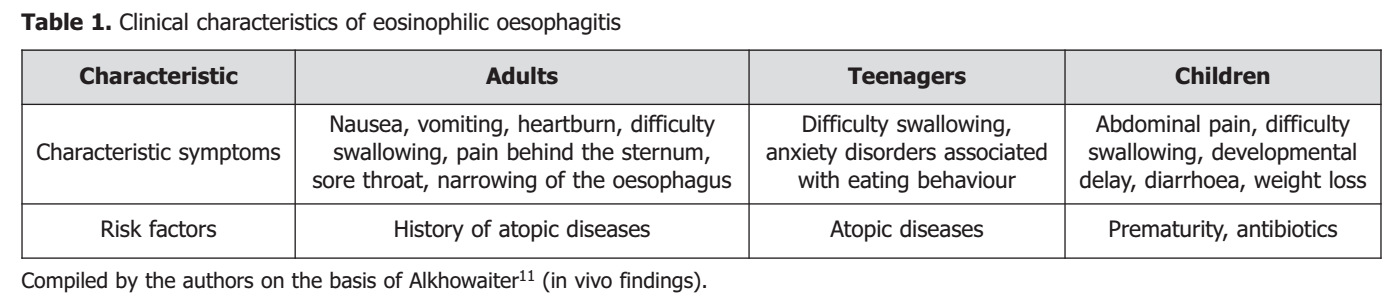
The difference in symptoms is explained by different pathophysiological changes in adults and children. It can also be attributed to the possibility of compensatory mechanisms in adults: slow eating, repeated chewing, cutting food into small pieces, and avoiding food that is uncomfortable to eat29. It is necessary to differentiate the disease from several other pathologies (oesophageal infections (fungal and viral), vasculitis, Morphine syndrome type 2, Netherton syndrome, atopic metabolic wasting syndrome, hypersensitivity due to medication, skin diseases (blistering), hyper immunoglobulin E (IgE) syndrome, Crohn’s disease with oesophageal involvement, celiac disease, oesophageal achalasia, eosinophilic gastritis, gastroesophageal reflux disease).
The I-SEE eosinophilic oesophagitis severity index is used to assess the severity of the disease. It includes three main categories: clinical picture and complications, inflammatory component, and fibrostenotic changes. The frequency of symptoms, the number and size of inflammatory signs (oedema, furrows, exudates, number of oesophageal eosinophils), and the presence of strictures are assessed (Figure 1). Each sign is assigned a score, with a total score of 1 to 15. Severity levels are determined depending on the number of points (1-6 mild, 7-14 moderate, 15 severe). The scale is relevant for long-term follow-up of patients.
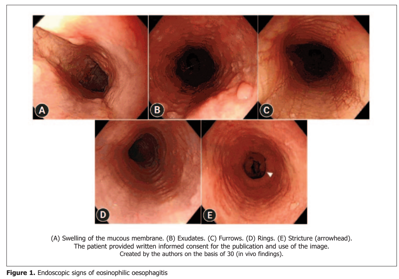
The diagnosis relies on evaluating oesophageal dysfunction symptoms, finding 15 or more eosinophils per high-power field in oesophageal biopsy samples, and confirming that eosinophilic inflammation is confined to the oesophagus. Esophagogastroduodenoscopy serves as the primary diagnostic tool, typically revealing white effusions and exudates, oesophageal rings, irregular luminal contours, strictures, erosions, and mucosal swelling with erythema. Wojas et al.4 noted that pathological signs of the lesion may be absent in 10% of adults and 30% of adults. Gonsalves and Aceves31 described that endoscopic sign in patients included normal appearance (30%), linear furrows (40%), oesophageal rings (14%), and white plaques (16%). It is worth noting that there are no pathognomonic endoscopic signs, and the severity of the disease does not correlate with symptoms, so a biopsy is always necessary. For diagnostic purposes, six sections are extracted from the proximal and distal regions of the oesophagus to enhance the examination’s efficacy. A positive result indicates an elevation in the quantity of intraepithelial eosinophils within the oesophagus, while they are absent in the stomach and duodenum. The advantages of endoscopy with biopsy are as follows: a proven system of evaluation and classification; the ability to predict histological remission and determine disease activity; and reliability in use both in individual cases and in systemic studies. Disadvantages are infrequent use in children; lack of consent among many patients; difficulties in implementation and risks of side effects in non-specialist healthcare facilities. Transnasal endoscopy is performed without sedation by passing a thin endoscope through the nose into the oesophagus. The advantage is that it can be performed on children as young as 6 years old. Barium oesophagography is more sensitive for the diagnosis of oesophageal stricture than conventional endoscopy. Radiography is useful for assessing the degree of fibrostenosis but is ineffective in diagnosing signs of inflammation and exudate32,33. Endoscopic ultrasound combined with high-resolution manometry increases the diagnostic benefit and facilitates the diagnosis of eosinophilic oesophagitis. This diagnosis enhances the comprehension of the pathomechanisms underlying oesophageal motility problems and the manifestation of clinical symptoms6. In the symptomatic course of eosinophilic oesophagitis, macroscopic changes in the upper gastrointestinal tract are detected. These changes often disappear with the use of steroids, and endoscopic ultrasound is a safe method for assessing the effectiveness of treatment. The use of endoscopic ultrasound in benign strictures allows visualising and analysing their length, location and level of damage. The oesophageal thread test involves the use of a capsule with a thread that is untangled in the oesophagus. The end of the thread is outside the patient’s mouth. The capsule remains in the oesophagus for 60 minutes, during which time the thread absorbs the contents of the oesophagus. The test has a sensitivity of 80% and a specificity of 77%4. Endoscopic ultrasound reveals thickening of the oesophageal walls. When using an endoscopic functional probe, a decrease in the compliance of the oesophageal wall was found. Wall remodelling occurs as a result of fibrosis of the mucosa and submucosa, and hypertrophy of smooth muscles. In adults, oesophageal diameter does not correlate with eosinophilic inflammation. At the same time, in children, the cross-sectional area of the oesophagus and its wall mobility correlate with eosinophilic inflammation and subepithelial fibrosis.34 Endoluminal functional lumen imaging sensor is a minimally invasive technology used to measure oesophageal distensibility. It is a new method that has a high potential for further study and use.
The diagnostic panel evaluates the expression of 96 genes that are affected in eosinophilic oesophagitis. It can be used to identify molecular phenotypes of eosinophilic oesophagitis. It is highly sensitive and specific. Moreover, 3 endotypes of eosinophilic oesophagitis have been identified: eosinophilic oesophagitis e1, eosinophilic oesophagitis e2 and eosinophilic oesophagitis e3. The initial endoscopic examination demonstrates a normal oesophageal appearance with mild histological alterations; there is a high expression of epithelial differentiation genes and a low expression of inflammatory and remodelling genes. The second kind is distinguished by pronounced histological alterations, elevated expression of inflammatory cytokine genes, and resistance to steroid treatment. The third type is distinguished by its commencement in adulthood, fibrostenotic alterations, and diminished expression of epithelial differentiation genes. This classification is important for a personalised approach to treatment. For instance, immunobiological drugs (anti-IL4Ra and anti-thymic stromal lymphopoietin) are effective in eosinophilic oesophagitis 2.
Treatment of eosinophilic oesophagitis
There are three main approaches to treating eosinophilic oesophagitis: dietary nutrition, medications and endoscopic oesophageal dilatation. Dietary nutrition includes the following models: elementary diet, elimination diet and empirical diet. An elementary diet is individually defined, well-balanced, and free of allergens. The efficiency is 92%, and the disadvantage is the bad taste and smell of the food. An elimination diet is prescribed based on the results of prick tests and tests for the level of specific IgE. The effectiveness is up to 50% in children and 30% in adults. The empirical diet involves the exclusion of the most allergenic foods (nuts, beans, wheat, eggs, fish and seafood). After 8 weeks of this diet, an endoscopic examination and biopsy are performed. Remission is reported in more than 70% of patients. The main disadvantage of this type of diet is the need to limit a large number of foods and conduct frequent endoscopic examinations. Therefore, a diet was developed to exclude the 2 most common allergens: wheat and cow milk. This approach ensured the disappearance of symptoms in 40% of patients.
Oesophageal dilatation is recommended in case of swallowing disorders and oesophageal stenosis up to 13 mm. The effectiveness in adults is up to 75%, and the risk of oesophageal perforation is less than 1%8. The number of dilatation sessions is less in the case of mucosal thickening and more in the case of submucosal lesions. Oesophageal dilation is aimed at reducing the manifestation of fibrostenotic components, while not affecting the level of eosinophilia. The advantage is a rapid reduction of symptoms with a low risk of relapse. The risk of side effects is low, Greenberg et al.35 reported that the incidence of perforation is 0-1%. Dilatation without concomitant drug therapy reduced the manifestations of dysphagia by 1 year and 3 months. At the same time, Schupack et al.36 described a decrease in the incidence of re-dilation in patients receiving maintenance drug therapy. Endoscopic oesophageal dilatation in combination with myotomy has shown high efficacy in the case of prolonged persistence of clinical symptoms, and ineffectiveness of steroid therapy against the background of fibrosis of the muscularis propria.
Proton pump inhibitors are used as part of drug treatment. They restore the epithelial barrier and reduce inflammation by decreasing eotaxin 3 and Th2 levels in the oesophageal mucosa; change the colonisation of the oesophageal microbiome increase IgE production; inhibit the expression of ICAM-1 and vascular cell adhesion molecules 1 enhance barrier function. In vitro, omeprazole blocks chromatin remodelling required for STAT6-mediated transcription of the eotaxin-3 gene. As a result of taking this group of drugs, the number of Bacteroidetes and Fusobacteria decreased, and the number of Firmicutes increased. Two-month use of omeprazole at a daily dose of 40 mg resulted in the disappearance of symptoms in 60% of patients and the disappearance of histological signs in 50% of patients. Adverse effects are infrequent and encompass diminished absorption of vitamin B12, reduced magnesium levels, and heightened risk of bone fractures. In vitro, JAKSTAT6 pathway inhibitors (ruxolitinib, leflunomide) reduce IL-13-induced eotaxin-3 production in oesophageal epithelial cells and fibroblasts, akin to proton pump inhibitors (PPIs). Tofacitinib elicited clinical and endoscopic remission in patients with resistant eosinophilic oesophagitis.
Topical steroids promote histological remission in more than 80% of patients and clinical remission in 70%. The mechanism of action is the inhibition of IL-13-dependent pathways and genes, which contributes to a decrease in eosinophils in the oesophagus, inhibition of epithelial tissue destruction, reduction in the number of T-lymphocytes and pro-inflammatory cytokines; the functions of the epithelial barrier are restored. Patients also showed a decrease in pro-fibrotic mediators: TGF-β1, FGF9 and IL-5. The advantage is low toxicity. Side effects are infrequent and include oesophageal candidiasis, adrenal cortex suppression, bone demineralisation and growth retardation. The treatment duration is 20-24 months. Factors associated with a lack of response to steroids include initial dilatation therapy prior to steroid use, oedema and/or decreased vascularity on endoscopic examination, hiatal hernia, and weight gain. Topical corticosteroids are metabolised in the liver, but cases of adrenal cortex suppression have been reported in patients with corticosteroid therapy. One-third of patients experienced side effects in the form of oesophageal candidiasis. Alternative means of delivering topical steroids are being developed to prolong the time of action of drugs on the gastric mucosa. The EsoCap system employs a specialised cup equipped with an applicator and a capsule containing a coiled polymer film. Upon swallowing the capsule, the film elongates and clings to the oesophagus. The gelatine capsule melts harmlessly in the oesophagus37,38. Systemic steroids reduce histological signs in more than 90% of patients, and symptoms disappear in 70%. The use of this group of drugs decreases the transcription of interleukin-13 and eosinophilic oesophagitis reduces the size of the eosinophilic infiltrate in 40-95% of patients and T-cell infiltration downregulates mast cell-associated genes and fibrosis and improves oesophageal motility. However, their main drawback is the high incidence of side effects, so this group of drugs should not be used routinely8,39,40. In children treated with corticosteroid therapy, a decrease in Actinobacillus was found. In patients with inactive eosinophilic oesophagitis, the number of Candida increased while taking steroids.
Dutta et al.41 described the drug Dupilumab, which is based on the monoclonal antibody IgG4, which binds the α-subunit of the heterodimeric IL-4 receptor. It is IL-4 and IL-13 that promote the release of eotaxin by oesophageal epithelial cells, which contributes to Th2 differentiation and the development of fibrotic changes. This product blocks the development of fibrotic changes and reduces the severity of the disease; it is the only biological product approved for the treatment of eosinophilic oesophagitis in people over 12 years of age. An injectable form of the drug was proposed with a frequency of administration of once every 7 days. Side effects include local drug reactions, nasopharyngitis fever, and in rare cases, conjunctivitis. Cendakimab is a humanised IgG1k monoclonal antibody that blocks IL-13 receptor binding, leading to improved histological and endoscopic outcomes after four months of treatment, though patients may experience headaches and nasopharyngitis. Mepolizumab, another IgG1k monoclonal antibody, binds circulating IL-5 to prevent receptor interaction and effectively reduces oesophageal eosinophilia, with injectable formulations showing superior efficacy. Benralizumab functions as an IgG1k monoclonal antibody-based therapeutic targeting the IL-5 receptor’s α-subunit, while tezepelumab operates as an IgG2λ monoclonal antibody that blocks thymic stromal lymphopoietin from binding its receptor. Lirentelimumab represents a humanised IgG1 antibody designed to target SIGLEC-8. This drug inhibits the activity of mast cells and reduces the number of eosinophils through apoptosis42. Omalizumab is a monoclonal antibody that attaches to free serum IgE, inhibiting its interaction with the high-affinity Fcε receptor. Natalizumab is a humanised monoclonal antibody that targets integrin α4 in integrins α4β1 and α4β7, resulting in the total resolution of eosinophilic oesophagitis, which does not respond to proton pump inhibitors and corticosteroids.
Prevention of eosinophilic oesophagitis
The aetiology of eosinophilic oesophagitis remains under investigation; nevertheless, many preventive strategies can mitigate the risk of disease onset and decrease the probability of sequelae. Individuals at risk should be closely monitored. If possible, breastfeeding should be preferred for newborns. Lyons et al.43 described that infant receive up to 8×10⁵ bacteria per day through daily breast milk intake. It is an important source of microbes for infants. Breastfeeding protects newborns from infectious diseases of the gastrointestinal tract, reduces the risk of chronic diseases, allergies, and irritable bowel syndrome, and improves the cognitive development of infants. This is due to its antimicrobial effect, prevention of pathogenic bacteria adhesion to the gastrointestinal epithelium and increased mucin production. The artificial creation of so-called “sterile” conditions in infancy is dangerous, as the lack of exposure to microbes in newborns increases the risk of allergic diseases. Aerts et al.44 and Markevych et al.45 note that exposure to a variety of microbes ensures the formation of a normal gastrointestinal microbiome and promotes the normal development of the immune system. Children who grew up in contact with domestic dogs attended kindergartens and had siblings have a lower risk of allergic diseases. There is a hypothesis of a relationship between polluted air and the development of allergies46-48. This is partly explained by the fact that exposure to external pollutants causes an inflammatory reaction involving cytokines, dendritic cells, basophils and Th2 cells in the skin, gastrointestinal tract and respiratory system (which are the first line of defence). This results in disruption of cell junctions and pathogen penetration. Krempski et al.49 and Vitte et al.50 note that the epithelium is similarly affected by chemicals in detergents.
Thus, the allergic origin of eosinophilic oesophagitis is indicated by several factors: allergic diseases in history; burdened family allergic history; peripheral eosinophilia; increased levels of total IgE; the presence of eosinophils in oesophageal samples; reduction of disease manifestations after elimination diet and steroid use. In the presence of genetic and systemic diseases, it is necessary to carefully monitor the condition of patients and, if symptoms that may indicate eosinophilic oesophagitis appear, immediately carry out several diagnostic measures. Early diagnosis and establishment of a diagnosis allow for better control of symptoms and avoidance of complications. It is worth noting that this is a chronic disease in which full recovery is not possible, but most patients experience symptom reduction with effective treatment. Maintenance therapy should be used to prevent a possible relapse.
CONCLUSIONS
Eosinophilic oesophagitis is a chronic Th2-mediated condition characterised by oesophageal dysfunction and eosinophilic inflammatory infiltrates. Cases have quadrupled over the past 25 years due to enhanced diagnostic methods and declining environmental quality in food, water, and air. The disease predominantly affects Caucasian males across all age groups, with risk factors including family or personal allergic disease history, early antibiotic exposure, and autoimmune or infectious conditions. The pathogenesis is still under study, but the mechanism of development is associated with immunological, genetic and environmental factors. The clinical picture differs depending on age. In infants, vomiting, general weakness, anxiety, refusal to eat, growth and developmental delays are observed against the background of acute inflammation. In adults, the symptoms are caused by the development of fibrosis, with men more often complaining of difficulty swallowing and women complaining of chest pain and heartburn. Diagnostic methods include esophagogastroduodenoscopy, biopsy, endoscopic ultrasound, and oesophageal thread test; the expression of genes damaged during eosinophilic oesophagitis is assessed. Several individually tailored diets are used to reduce symptoms. There is currently no etiological treatment. Therefore, symptomatic therapy is used: proton pump inhibitors, and local and systemic steroids. New products have been introduced that allow to increase in the local time of steroid exposure (e.g., EsoCup). In cases of severe stenosis, oesophageal dilatation is used. The method is highly effective, and safe and significantly reduces the risk of relapse. It is most effective in combination with concomitant steroid therapy.
The limitations of the present study are the insufficiently studied aetiology and pathogenesis of eosinophilic oesophagitis. Also, the study’s methodological limitations include the reliance on two reviewers for screening and selection, which may introduce bias, and the potential for variability in the quality of evidence across the included study designs. Therefore, further study of the above issues is needed to develop effective treatments for eosinophilic oesophagitis.
REFERENCES
- Wąsik J, Małecka-Wojciesko E. Eosinophilic esophagitis what do we know so far? J Clin Med. 2023;12(6):2259. doi: 10.3390/jcm12062259.
- Kanikowska A, Hryhorowicz S, Rychter AM, Kucharski MA, Zawada A, Iwanik K, Eder P, Słomski R, Dobrowolska A, Krela-Kaźmierczak I. Immunogenetic, molecular and microbiotic determinants of eosinophilic esophagitis and clinical practice – A new perspective of an old disease. Int J Mol Sci. 2021;22(19):10830. doi: 10.3390/ijms221910830.
- Savarino EV, Barbara G, Bilò MB, De Bortoli N, Di Sabatino A, Oliva S, Penagini R, Racca F, Tortora A, Rumi F, Cicchetti A. Eosinophilic esophagitis in adults and adolescents: Epidemiology, diagnostic challenges, and management strategies for a type 2 inflammatory disease. Ther Adv Gastroenterol. 2024;17. doi: 10.1177/17562848241249570.
- Wojas O, Żalikowska-Gardocka M, Krzych-Fałta E, Samoliński B, Przybyłkowski A. Eosinophilic esophagitis: An interdisciplinary clinical problem. Postep Dermatol Alergol. 2021;38(1):36-42. doi: 10.5114/ada.2019.87237.
- Muir A, Falk GW. Eosinophilic esophagitis: A review. JAMA. 2022;326(13):1310-1318. doi: 10.1001/jama.2021.14920.
- Zhang X, Zhang N, Wang Z. Eosinophilic esophagitis and esophageal microbiota. Front Cell Infect Microbiol. 2023;13:1206343. doi: 10.3389/fcimb.2023.1206343.
- Pytrus T, Akutko K, Kofla-Dłubacz A. Endoscopic ultrasonography in children with eosinophilic esophagitis – A review. Pediatr Rep. 2022;14(1):13-19. doi: 10.3390/pediatric14010003.
- Sarbinowska J, Wiatrak B, Waśko-Czopnik D. Searching for noninvasive predictors of the diagnosis and monitoring of eosinophilic esophagitis – The importance of biomarkers of the inflammatory reaction involving eosinophils. Biomolecules. 2021;11(6):890. doi: 10.3390/biom11060890.
- Kuźmiński A, Przybyszewski M, Przybyszewska J, Bartuzi Z. Eosinophilic esophagitis. What diet? How to treat it? The point of view of a gastroenterologist working in a department of allergology. Przegl Gastroenterol. 2021;16(4):318-323. doi: 10.5114/pg.2021.105163.
- Główczewski A, Krogulska A. Formulations of topical steroids in eosinophilic esophagitis – Current treatment and emerging possibilities. J Clin Med. 2022;11(5):1454. doi: 10.3390/jcm11051454.
- Alkhowaiter S. Eosinophilic esophagitis. Saudi Med J. 2023;44(7): 640-646. doi: 10.15537/smj.2023.44.7.20220812.
- Chang Y, Haller E, Dellon E. Dietary management of eosinophilic esophagitis: Man versus food or food versus man? Gastroenterol Clin North Am. 2021;50(1):59-75. doi: 10.1016/j.gtc.2020.10.009.
- Loi R, Ceulemans M, Wauters L, Vanuytsel T. An update on eosinophilic esophagitis. Acta Gastroenterol Belg. 2023;86(4):533-542. doi: 10.51821/86.4.12001.
- Votto M, Filippo M, Caimmi S, Indolfi C, Raffaele A, Tosca MA, Marseglia GL, Licari A. A practical update on pediatric eosinophilic esophagitis. Children (Basel). 2023;10(10):1620. doi: 10.3390/children10101620.
- Khokhar D, Marella S, Idelman G, Chang JW, Chehade M, Hogan SP. Eosinophilic esophagitis: Immune mechanisms and therapeutic targets. Clin Exp Allergy. 2022;52(10):1142-1156. doi: 10.1111/cea.14196.
- Talley N, Burns G, Hoedt E, Duncanson K, Keely S. Beyond eosinophilic esophagitis: Eosinophils in gastrointestinal disease-new insights, “new” diseases. J Can Assoc Gastroenterol. 2023; 6(6):199-211. doi: 10.1093/jcag/gwad046.
- Massironi S, Mulinacci G, Gallo C, Elvevi A, Danese S, Invernizzi P, Vespa E. Mechanistic insights into eosinophilic esophagitis: Therapies targeting pathophysiological mechanisms. Cells. 2023; 12(20):2473. doi: 10.3390/cells12202473.
- Muir A, Karakasheva T, Whelan K. Epithelial-fibroblast crosstalk in eosinophilic esophagitis. Cell Mol Gastroenterol Hepatol. 2024; 17(5):713-718. doi: 10.1016/j.jcmgh.2024.01.020.
- Gupta M, Grinman M. Diagnosis and management of eosinophilic esophagitis. CMAJ. 2024;196(4):E121-E128. doi: 10.1503/cmaj.230378.
- de Bortoli N, Visaggi P, Penagini R, Annibale B, Baiano Svizzero F, Barbara G, Bartolo O, Battaglia E, Di Sabatino A, De Angelis P, Docimo L, Frazzoni M, Furnari M, Iori AV, Marabotto E, Marasco G, Mauro A, Oliva S, Savarino EV. The 1st EoETALY consensus on the diagnosis and management of eosinophilic esophagitis – Definition, clinical presentation and diagnosis. Dig Liver Dis. 2024;56(6):951-963. doi: 10.1016/j.dld.2024.02.005.
- Cavalli E, Brusaferro A, Pieri E, Cozzali R, Farinelli E, De’ Angelis GL, Esposito S. Eosinophilic esophagitis in children: doubts and future perspectives. J Transl Med. 2019;17(1):262. doi: 10.1186/s12967-019-2014-0.
- O’Shea K, Rochman M, Shoda T, Zimmermann N, Caldwell J, Rothenberg ME. Eosinophilic esophagitis with extremely high esophageal eosinophil counts. J Allergy Clin Immunol. 2021; 147(1):409-412. doi: 10.1016/j.jaci.2020.05.045.
- Bolton S, Kagalwalla A, Arva N, Wang MY, Amsden K, Melin-Aldana H, Dellon ES, Bryce PJ, Wershil BK, Wechsler JB. Mast cell infiltration is associated with persistent symptoms and endoscopic abnormalities despite resolution of eosinophilia in pediatric eosinophilic esophagitis. Am J Gastroenterol. 2020;115(2):224-233. doi: 10.14309/ajg.0000000000000474.
- Young E, Philpott H. Pathophysiology of dysphagia in eosinophilic esophagitis: Causes, consequences, and management. Dig Dis Sci. 2022;67(4):1101-1115. doi: 10.1007/s10620-022-07419-6.
- Alsohaibani F, Peedikayil M, Alzahrani M, Azzam NA, Almadi MA, Dellon ES, Al-Hussaini AA. Eosinophilic esophagitis: Current concepts in diagnosis and management. Saudi J Gastroenterol. 2024; 30(4):210-227. doi: 10.4103/sjg.sjg_50_24.
- Yang EJ, Jung KW. Role of endoscopy in eosinophilic esophagitis. Clin Endosc. 2024;48(5):10-13. doi: 10.5946/ce.2024.023.
- Hiremath G, Yazdian A, Onuh I, Willey J, Choksi Y. Race and gender influences the presentation of eosinophilic esophagitis. Dysphagia. 2023;38(6):1511-1518. doi: 10.1007/s00455-023-10577-y.
- Larsson H, Strobel M, Perez-Guagnelli E. Emotional journey of patients with eosinophilic esophagitis. Adv Ther. 2023;40(12):5254-5270. doi: 10.1007/s12325-023-02678-9.
- Laserna-Mendieta EJ, Navarro P, Casabona-Francés S, Savarino EV, Amorena E, Pérez-Martínez I, Guagnozzi D, Blas-Jhon L, Betoré E, Guardiola-Arévalo A, Pellegatta G, Krarup AL, Perello A, Barrio J, Gutiérrez-Junquera C, Teruel Sánchez-Vegazo C, Fernández-Fernández S, Naves JE, Oliva S, Rodríguez-Oballe JA. Swallowed topical corticosteroids for eosinophilic esophagitis: Utilization and real-world efficacy from the EoE CONNECT registry. United Eur Gastroenterol J. 2024;12(5):585-595. doi: 10.1002/ueg2.12533.
- Yang EJ, Jung KW. Role of endoscopy in eosinophilic esophagitis. Clin Endosc. 2024;48(5):10-13. doi: 10.5946/ce.2024.023.
- Gonsalves N, Aceves S. Diagnosis and treatment of eosinophilic esophagitis. J Allergy Clin Immunol. 2021;145(1):1-7. doi: 10.1016/j.jaci.2019.11.011.
- Suzuki Y, Ochiai Y, Hosoi A, Okamura T, Hayasaka J, Mitsunaga Y, Tanaka M, Odagiri H, Nomura K, Yamashita S, Matsui A, Kikuchi D, Ohashi K, Hoteya S. Mucosal and submucosal thickening of esophageal wall is a promising factor in the development of symptoms in eosinophilic esophagitis. Gut Liver. 2024;18(1):50-59. doi: 10.5009/gnl220490.
- Visaggi P, Ghisa M, Vespa E, Barchi A, Mari A, Pasta A, Marabotto E, de Bortoli N, Savarino EV. Optimal assessment, treatment, and monitoring of adults with eosinophilic esophagitis: Strategies to improve outcomes. ImmunoTargets Ther. 2024;13:367-383. doi: 10.2147/ITT.S276869.
- Samanta A, Poddar U, Kumari N, Sen Sarma M, Srivastava A, Mishra P. Eosinophilic esophagitis in children: A cross-sectional study from a tertiary care center. JGH Open. 2023;8(1):e13024. doi: 10.1002/jgh3.13024.
- Greenberg S, Chang N, Corder R, Reed C, Eluri S, Dellon ES. Dilation-predominant approach versus routine care in patients with difficult-to-treat eosinophilic esophagitis: A retrospective comparison. Endoscopy. 2022;54(3):243-250. doi: 10.1055/a-1493-5627.
- Schupack DA, Ravi K, Geno DM, Pierce K, Mara K, Katzka DA, Alexander JA. Effect of maintenance therapy for eosinophilic esophagitis on need for recurrent dilation. Dig Dis Sci. 2021; 66(2):503-510. doi: 10.1007/s10620-020-06192-8.
- Petrousis G, Retsas P, Ignatova S, Karapiperis D. Treatment-refractory eosinophilic esophagitis successfully managed with benralizumab: A case presentation and literature review. Rom J Intern Med. 2024;62(3):356-361. doi: 10.2478/rjim-2024-0021.
- Kai H, Cao L, Qi L, Zhu J, Xu H, Fang J. Progress in clinical treatment and nursing of pediatric eosinophilic esophagitis. Minerva Medica. 2024;115(2):243-245. doi: 10.23736/S0026-4806.23.08626-3.
- Sia T, Miller A, Bacchus L, Young J, Narayan AP, Solecki R, Fu J, Jiang Y, Khuda R, Liu S, Love K, Mallik S, Matmatte AS, McDonald P, Telukunta T, Roby A, Shami S, Zheng M, Headen M, Leung J. Dupilumab improves clinical and histologic features of eosinophilic esophagitis prior to 12 weeks of treatment. Clin Transl Allergy. 2024;14(1):e12333. doi: 10.1002/clt2.12333.
- Lombardi C, Comberiati P, Ridolo E, Cottini M. Anti-IL-5 pathway agents in eosinophilic-associated disorders across the lifespan. Drugs. 2024;84(6):661-684. doi: 10.1007/s40265-024-02037-0.
- Dutta P, Shah-Riar P, Bushra S. Recent trends in the management of eosinophilic esophagitis: A systematic review. Cureus. 2023; 15(8):e43221. doi: 10.7759/cureus.43221.
- Dellon ES, Khoury P, Muir AB, Liacouras CA, Safroneeva E, Atkins D, Collins MH, Gonsalves N, Falk GW, Spergel JM, Hirano I, Chehade M, Schoepfer AM, Menard-Katcher C, Katzka DA, Bonis PA, Bredenoord AJ, Geng B, Jensen ET, Pesek RD, Aceves SS. A clinical severity index for eosinophilic esophagitis: development, consensus, and future directions. Gastroenterology. 2023;163(1): 59-76. doi: 10.1053/j.gastro.2022.03.025.
- Lyons K, Ryan C, Dempsey E, Ross P, Stanton C. Breast milk, a source of beneficial microbes and associated benefits for infant health. Nutrients. 2020;12(4):1039. doi: 10.3390/nu12041039.
- Aerts R, Honnay O, Nieuwenhuyse A. Biodiversity and human health: Mechanisms and evidence of the positive health effects of diversity in nature and green spaces. Br Med Bull. 2018;127(1): 5-22. doi: 10.1093/bmb/ldy021.
- Markevych I, Schoierer J, Hartig T, Chudnovsky A, Hystad P, Dzhambov AM, de Vries S, Triguero-Mas M, Brauer M, Nieuwenhuijsen MJ, Lupp G, Richardson EA, Astell-Burt T, Dimitrova D, Feng X, Sadeh M, Standl M, Heinrich J, Fuertes E. Exploring pathways linking greenspace to health: Theoretical and methodological guidance. Environ Res. 2017;158:301-317. doi: 10.1016/j.envres.2017.06.028.
- Street M, Shulhai AM, Rotondo R, Giannì G, Caffarelli C. Current knowledge on the effects of environmental contaminants in early life nutrition. Front Nutr. 2023;10:1120293. doi: 10.3389/fnut.2023.1120293.
- Boonpiyathad T, Sözener Z, Satitsuksanoa P, Akdis CA. Immunologic mechanisms in asthma. Semin Immunol. 2019;46:101333. doi: 10.1016/j.smim.2019.101333.
- Mabotja K, Onselen A, Gordon R. Infant feeding knowledge, attitudes and practices of HIV-positive breastfeeding mothers. Health SA. 2024;29:2617. doi: 10.4102/hsag.v29i0.2617.
- Krempski J, Dant C, Nadeau K. The origins of allergy from a systems approach. Ann Allergy Asthma Immunol. 2020;125(5):507-516. doi: 10.1016/j.anai.2020.07.013.
- Vitte J, Vibhushan S, Bratti M, Montero-Hernandez J, Blank U. Allergy, anaphylaxis, and nonallergic hypersensitivity: IgE, mast cells, and beyond. Med Princ Pract. 2022;31(6):501-515. doi: 10.1159/000527481.
Explorar
Artículos
Volúmenes
Sobre la revista
Más leídos
Consumo de bebidas energizantes y efectos adversos en la salud de estudiantes universitarios
Nutrición Clínica y Dietética Hospitalaria
Uso de redes sociales y riesgo de trastornos de conducta alimentaria en adolescentes mujeres de un colegio en Lima, Perú
Nutrición Clínica y Dietética Hospitalaria
Consumo de bebidas energizantes en estudiantes universitarios y sus factores asociados.
Nutrición Clínica y Dietética Hospitalaria
Microbiota intestinal y eje intestino-cerebro: una revisión de su interacción con el sistema nervioso central
Nutrición Clínica y Dietética Hospitalaria
Factores asociados a la diabetes mellitus tipo 2 de la población adulta peruana.
Nutrición Clínica y Dietética Hospitalaria